Dr. David Tzou, Dr. Thomas Chi, and their colleagues from the UCSF Departments of Radiology and Urology have recently published a case report regarding a new application of contrast-enhanced ultrasound for a diagnosis of kidney cancer recurrence .
Patients with chronic renal impairment and a history of renal cell carcinoma represent a challenging patient population with respect to surveillance management. These patients are often poor candidates for iodinated or gadolinium contrast administration commonly used in computed tomography (CT scan) or magnetic resonance imaging (MRI) due to a high risk of contrast toxicity. The authors report a case of an elderly patient with a solitary left kidney who underwent left partial nephrectomy for renal cell carcinoma over five years ago. The patient then developed gross hematuria and ultimately benefitted from the use of contrast-enhanced ultrasound compared to CT and MRI to diagnose a recurrent mass. This case report demonstrates a new role for contrast-enhanced ultrasound in the surveillance management of patients with renal failure and renal cell carcinoma.
Case Report: Contrast Enhanced Ultrasound Detects Recurrent Renal Cell Carcinoma in the Setting of Chronic Renal Insufficiency. Tzou DT, Weinstein S, Usawachintachit M, Mongan J, Greene KL, Chi T. Clin Genitourin Cancer. 2016. http://dx.doi.org/10.1016/j.clgc.2016.12.010 Read more...
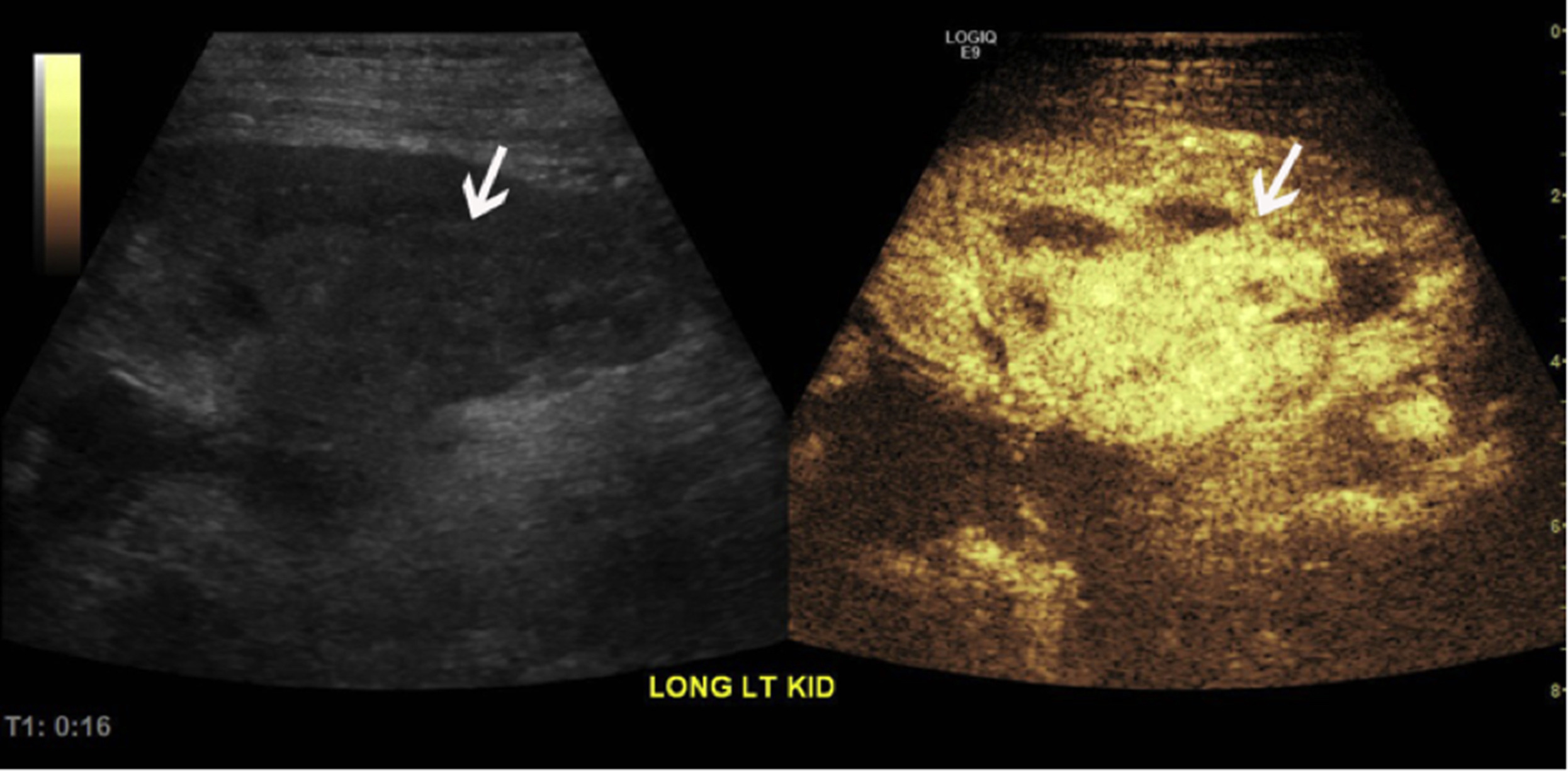
On the left, the gray-scale ultrasound image demonstrates a possible vague area of soft tissue fullness that is incompletely characterized (white arrow). The corresponding contrast-enhanced ultrasound image on the right confirms a discrete mass with marked hypervascularity (white arrow) compared with adjacent renal parenchyma consistent with a hypervascular mass.